top of page


Removal of Myxoma
Removal of Myxoma
Masses are not commonly found within the heart. But masses within the heart can require surgical attention.
The most common mass in the heart is a mass from somewhere else, which is called a secondary tumor. The #1 mass in the heart is a cancer mass (from somewhere else).
A mass that arises within the heart itself is called a primary tumor. Primary heart tumors are rare.
The most common primary heart mass is a myxoma. A myxoma is not cancerous.
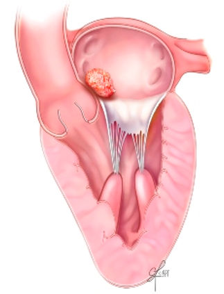
A myxoma most commonly arises within the atria of the heart. The left atrium is the classic location. But a myxoma can arise from almost any structure within the heart.
A patient may not know he or she has a mass within the heart, especially if the mass is small.
A mass can become large and begin to block the bloodflow through the heart. Or the mass can interfere with the opening and closing of a heart valve.
Or a piece of the mass can break off, which can travel to the brain and cause a stroke.
A patient may experience shortness of breath or ankle swelling or chest pain if a mass grows to significant size and begins to affect the heart function. Some masses may cause fatigue or tiredness or fever.
A myxoma is usually diagnosed by an echocardiogram, a fancy ultrasound.
In this case, the myxoma is large and moves in and out of the mitral valve with each heartbeat.
Typically, a myxoma should be removed promptly upon diagnosis, in order to minimize the risk of stroke or heart failure.
If a myxoma is identified and needs to be removed, there are a couple of options for surgical approach:

STERNOTOMY

MINIMALLY-INVASIVE

A sternotomy can be used to perform removal of myxoma.
The patient is put to sleep with a general anesthetic.
An incision is made thru the breast-bone and the heart is exposed.
The heart and lung machine is used, so that the heart can be stopped.
Once the heart is stopped, an incision is made to access the myxoma.
The myxoma is removed and sent to pathology for evaluation.
Patients are usually in the hospital 4-5 days after a sternotomy.

A minimally-invasive method can often be used for removal of myxoma.
The patient is put to sleep with a general anesthetic.
A minimally-invasive approach usually takes 1-2 hours to perform.
Dr. Pool has taken out many myxomas with a minimally-invasive approach.
An incision is made in the right chest, between the ribs. The breastbone is not cut.
A small incision is also made in the groin.
Patients are usually in the hospital 4-5 days after a minimally-invasive removal of myxoma.
The heart and lung machine is used, the heart is stopped, and the myxoma is removed.
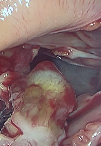
A myxoma often has similarity in appearance and consistency of jello.

Aortic Stenosis
bottom of page