top of page


Plication of Diaphragm
Plication of Diaphragm
The thoracic diaphragm is a large, thin muscle which separates the chest from the abdomen.

The diaphragm is made of skeletal muscle. This means that it can be voluntarily contracted. Of course, normal, comfortable breathing does not require conscious effort for most people.
The diaphragm contracts, increasing space in the thorax and allowing air to passively enter the lungs, since the pressure is lower in the lungs than in the atmosphere.
The diaphragm has two domes, right and left.
The diaphragm is innervated by the phrenic nerve. The phrenic nerve arises from the neck. To be clear, there are two phrenic nerves, one on right and one on left. The phrenic nerves travel down the middle of the chest near the heart and provide input to the diaphragm.
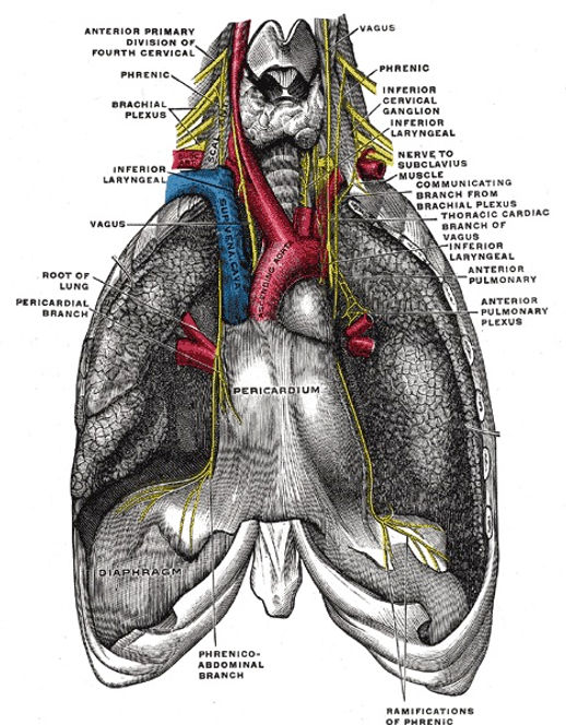
The reason this anatomy is important is that if one of the phrenic nerves is injured or becomes damaged anywhere along its course from the neck to the bottom of the chest, the diaphragm may not contract properly.
If the diaphragm does not contract, it is said to be paralyzed.
When the diaphragm is paralyzed, the patient may experience shortness of breath. This shortness of breath may occur even when the patient is at rest.
_chest_radiog.jpg)
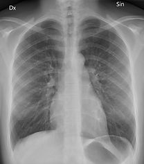
Normal Chest X-ray

Paralyzed Right Diaphragm
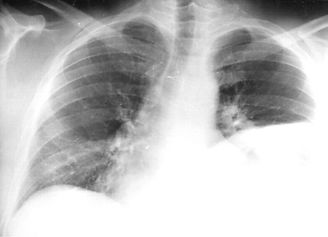
Paralyzed Left Diaphragm
A paralyzed diaphragm usually results in a chest x-ray which shows elevation of the diaphragm. The diaphragm becomes flimsy, stretched out. The lung is compressed and the air does not move as efficiently.
If the damage to the phrenic nerve is not permanent, some diaphragmatic function may return. If the damage is permanent and the patient suffers from shortness of breath, a plication of the diaphragm may help.
The idea behind plication of the diaphragm is that the flabby, stretched out dome of the diaphragm can be plicated, or folded in upon itself, to make it snug.

Stitches are put in that will gather up the flabby diaphragm and cinch it in.
This allows the lung to expand more fully and reduces the patient's shortness of breath.
Dr. Pool prefers to perform plication of diaphragm with a robotic approach.
With the patient under a general anesthetic, 3 or 4 small incisions (less than an inch each) are made in the side of the chest, between the ribs.
Sutures are placed in the diaphragm, gathering in the flabby portions. The sutures are tied, making the diaphragm snug.
A chest tube - which is like a straw - is placed in the chest.
If both sides of the diaphragm are paralyzed then the plication may be done on both sides.
Patients do not usually need to go to the ICU after operation.
Patients are usually in the hospital 1-2 days after a robotic plication of diaphragm.
A robotic plication usually takes 1-2 hours to perform.
Plication video
bottom of page